



Tomland Medical Movable Knee Joint Model with Ligaments & Menisci
$69.95
In stock (can be backordered)

Clinical Pricing Available!
The Physio Store is offering clinical pricing for all businesses / institutions in healthcare, healthcare professionals/students and fitness industry professionals.
We highly recommend you to sign up for our clinical pricing program, which will provide you with discounts on most of our products. We will also send out exclusive monthly specials, new ideas and new products on the market. Our company values the hard work and dedication of healthcare professionals and express gratitude by offering you special pricing and monthly deals.
Close
Proudly Canadian-Owned and Operated Company!
This product comes from a Proudly Canadian Company, with its owners and headquarters based right here in Canada. As a locally owned business, we are committed to providing high-quality products while supporting our community and economy. By choosing this product, you are supporting a Canadian company dedicated to excellence, customer care, and innovation. Thank you for supporting local!
Close

Manufacturer Warranty
Warranty Coverage
Return Policy
30-Day Return Window. Returns are accepted within 30 days of delivery for items that remain new and unopened.
Unopened condition required. Security seals must remain intact and packaging must not be opened. Items must be returned in original, unused condition for full product credit.
All law labels, identification labels, manuals, and original packaging must remain attached and undamaged. Please pack returns carefully in a secondary shipping box to prevent damage in transit.
If an item is returned opened, used, or not in resellable condition, the return will not be accepted. You will be contacted regarding return shipping fees to have the item shipped back to you.
If we do not receive instructions regarding return shipment within 30 days, you authorize us to dispose of the item on your behalf.
International orders are final sale. Orders shipped outside of Canada are not eligible for return or exchange.
For complete eligibility requirements and return instructions, please review our Return & Exchange Policy.

Tomland Medical Movable Knee Joint Model with Ligaments & Menisci
The Tomland Medical Movable Knee Joint Model is an economical anatomy model designed for clinics, classrooms and patient education. It shows the normal knee joint with the femur, tibia, fibula, patella, major knee ligaments and medial and lateral menisci.
This model is especially useful for healthcare professionals who want to explain how the knee bends and straightens. Because the model can flex and extend, patients and students can better understand how the knee joint moves and how the ligaments, menisci and patella relate to one another.
Movable Knee Anatomy Teaching Model
- Demonstrates normal knee joint anatomy
- Flexes and extends to show basic knee movement
- Shows the femur, tibia, fibula and patella
- Includes visible ACL, PCL, MCL, LCL and patellar ligament
- Shows the medial and lateral menisci
- Patella can be turned to view the posterior surface and facets
- Mounted on a display base for teaching and presentation
Why Use a Movable Knee Joint Model?
Knee injuries and knee pain can be difficult to explain using words alone. A physical model allows clinicians to point directly to the ligaments, menisci, patella and joint surfaces being discussed.
In addition, the movable design helps demonstrate knee flexion and extension. This can make patient conversations clearer when discussing general knee mechanics, ligament injuries, meniscus problems, patellar tracking, arthritis, rehabilitation exercises or post-injury education.
Helpful For Demonstrating
- Normal knee flexion and extension
- Basic knee joint structure
- ACL, PCL, MCL and LCL location
- Medial and lateral meniscus position
- Patella position and posterior patellar surface
- Relationship between the femur, tibia and fibula
- General knee injury and rehabilitation education
Who Is It For?
Common Users
- Physiotherapy clinics
- Chiropractic clinics
- Massage therapy clinics and schools
- Athletic therapy and sports medicine clinics
- Kinesiology and rehabilitation programs
- Medical, nursing and health-science classrooms
- Personal trainers and movement educators
Product Features
- Economical normal knee joint anatomy model
- Movable design allows flexion and extension demonstration
- Bendable artificial ligaments for visual education
- Shows the medial and lateral femoral condyles
- Shows the medial and lateral tibial condyles
- Includes patella, tibia, femur and fibula
- Includes visible collateral and cruciate ligaments
- Includes visible medial and lateral menisci
- Made from PVC material according to manufacturer specifications
- Mounted on a base for display and demonstration
Product Specifications
| Brand | Tomland Medical |
|---|---|
| Product Type | Movable knee joint anatomy model |
| Anatomy Shown | Femur, tibia, fibula, patella, ligaments and menisci |
| Movement | Flexion and extension demonstration |
| Visible Ligaments | ACL, PCL, MCL, LCL and patellar ligament |
| Visible Menisci | Medial and lateral meniscus |
| Patella | Can be turned to view the posterior surface and facets |
| Material | PVC according to manufacturer specifications |
| Display | Mounted on base |
| Use | Educational and patient demonstration use |
| Ideal For | Clinics, classrooms, training programs and patient education |
Good to Know
- This is a normal knee joint model intended for general anatomy education.
- It is best suited for patient demonstrations, classroom teaching and basic anatomy review.
- It is not intended for diagnosis, surgical planning, implant fitting or advanced biomechanical testing.
- Exact model details, colour and finish may vary slightly between production batches.
Tomland Medical Anatomy Models
Warranty Coverage
Frequently Asked Questions
What is the Tomland Medical Movable Knee Joint Model used for?
This model is used for knee anatomy education, patient demonstrations and classroom teaching. It helps show the femur, tibia, fibula, patella, ligaments and menisci in a clear visual format.
Does this knee joint model move?
Yes. The model can flex and extend, which makes it useful for demonstrating basic knee movement during patient education or anatomy teaching.
Which knee ligaments are shown on this model?
This model shows the ACL, PCL, MCL, LCL and patellar ligament, making it useful for explaining common knee ligament anatomy.
Does the model show the meniscus?
Yes. The model shows both the medial meniscus and lateral meniscus, which can help explain meniscus location and general knee structure.
Does this model include the patella?
Yes. The model includes the patella, and the patella can be turned to view the posterior surface and patellar facets.
Is this knee model suitable for physiotherapy clinics?
Yes. Physiotherapy clinics can use this model to explain knee injuries, ligament location, meniscus position, patellar movement and general rehabilitation concepts.
Can chiropractors, massage therapists or athletic therapists use this model?
Yes. This model is useful for many healthcare and movement professionals who explain knee anatomy or knee movement to patients and students.
Is this a normal knee joint model?
Yes. This is a normal knee joint anatomy model. It is designed to show general knee structure and movement rather than a specific disease or surgical condition.
What bones are shown on this knee model?
The model shows the femur, tibia, fibula and patella, which are the main bony structures commonly reviewed when explaining knee anatomy.
Is this model useful for explaining ACL or meniscus injuries?
Yes. While it does not show an injured knee, it can help explain where the ACL, PCL and menisci are located, which can make injury education easier to understand.
Is this knee joint model mounted on a base?
Yes. The model is mounted on a display base, making it easy to use in a clinic room, classroom or teaching area.
Is this model intended for diagnosis or surgical planning?
No. This model is intended for education and visual demonstration only. It should not be used for diagnosis, surgical planning, implant fitting or biomechanical testing.
| Weight | 870 g |
|---|---|
| Dimensions | 30 × 11 × 11 cm |
| HS Code | 9023.00.00.00 |
| Country of Origin | China |
| Availability |
Now available in-store at 22635 Komoka Road Komoka N0L1R0 Powered With Oliver Pos |
|---|
What is the Tomland Medical Movable Knee Joint Model used for?
This model is used for knee anatomy education, patient demonstrations and classroom teaching. It helps show the femur, tibia, fibula, patella, ligaments and menisci in a clear visual format.
Does this knee joint model move?
Yes. The model can flex and extend, which makes it useful for demonstrating basic knee movement during patient education or anatomy teaching.
Which knee ligaments are shown on this model?
This model shows the ACL, PCL, MCL, LCL and patellar ligament, making it useful for explaining common knee ligament anatomy.
Does the model show the meniscus?
Yes. The model shows both the medial meniscus and lateral meniscus, which can help explain meniscus location and general knee structure.
Does this model include the patella?
Yes. The model includes the patella, and the patella can be turned to view the posterior surface and patellar facets.
Is this knee model suitable for physiotherapy clinics?
Yes. Physiotherapy clinics can use this model to explain knee injuries, ligament location, meniscus position, patellar movement and general rehabilitation concepts.
Can chiropractors, massage therapists or athletic therapists use this model?
Yes. This model is useful for many healthcare and movement professionals who explain knee anatomy or knee movement to patients and students.
Is this a normal knee joint model?
Yes. This is a normal knee joint anatomy model. It is designed to show general knee structure and movement rather than a specific disease or surgical condition.
What bones are shown on this knee model?
The model shows the femur, tibia, fibula and patella, which are the main bony structures commonly reviewed when explaining knee anatomy.
Is this model useful for explaining ACL or meniscus injuries?
Yes. While it does not show an injured knee, it can help explain where the ACL, PCL and menisci are located, which can make injury education easier to understand.
Is this knee joint model mounted on a base?
Yes. The model is mounted on a display base, making it easy to use in a clinic room, classroom or teaching area.
Is this model intended for diagnosis or surgical planning?
No. This model is intended for education and visual demonstration only. It should not be used for diagnosis, surgical planning, implant fitting or biomechanical testing.
You may also like…


Related products

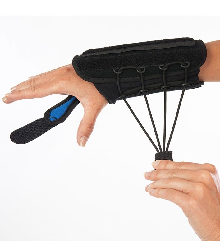
ProCare Quick-Fit II Wrist Brace
In Stock
$45.95

DonJoy Lateral J Patella Knee Brace
In Stock
$134.95
Elasto-Gel Shoulder Sleeve
In Stock
Price range: $129.95 through $144.95
Serola Sacroiliac Belt – version 2
In Stock
$79.95
 SUPPORT SMALL CANADIAN BUSINESS


Reviews
There are no reviews yet