



TomLand Medical 4-Stage Knee Osteoarthritis Model
$79.95
In stock (can be backordered)

Clinical Pricing Available!
The Physio Store is offering clinical pricing for all businesses / institutions in healthcare, healthcare professionals/students and fitness industry professionals.
We highly recommend you to sign up for our clinical pricing program, which will provide you with discounts on most of our products. We will also send out exclusive monthly specials, new ideas and new products on the market. Our company values the hard work and dedication of healthcare professionals and express gratitude by offering you special pricing and monthly deals.
Close

Manufacturer Warranty
Return Policy
Final Sale Item. This product is not eligible for return, refund, or exchange once purchased.
High-Risk Medical Devices & Hygienic Products
Certain items are designated as Final Sale due to their medical, personal-use, hygienic, or electrical nature. These products cannot be restocked or resold once opened or shipped.
This includes (but is not limited to):
- Electrotherapy devices (TENS, EMS, IFC units)
- Items that come into contact with skin or bodily areas
- Personal-use rehabilitation and pain management devices
- Electrical medical equipment
For health, safety, and product integrity reasons, these items are non-returnable regardless of usage.
Clearance & Special Order Items
Products marked as Clearance, Special Order, or End-of-Line are also Final Sale and not eligible for return due to limited inventory availability.
Exceptions apply only for defective, damaged, or incorrectly shipped items reported within 5 days of delivery.
By completing your purchase, you acknowledge and agree to these Final Sale terms prior to checkout.
For full details, please review our Return & Exchange Policy.

TomLand Medical 4-Stage Knee Osteoarthritis Model
The TomLand Medical 4-Stage Knee Osteoarthritis Model is a practical teaching model for explaining knee joint wear over time. It shows four comparison stages: a normal knee joint, early degeneration, moderate degeneration and severe degeneration.
This model helps clinicians, educators and students explain common knee arthritis changes. For example, it can help show cartilage wear, joint-space narrowing, osteophyte formation, meniscus deterioration and more advanced joint changes.
Helpful for Knee Osteoarthritis Education
- Shows a normal knee joint plus early, moderate and severe degeneration
- Helps explain cartilage loss, joint-space changes and osteophyte formation
- Shows meniscus deterioration and progressive knee joint wear
- Supports patient education in clinics, classrooms and training programs
- Provides a clear visual comparison between healthy and arthritic knee changes
Why Use a 4-Stage Knee Osteoarthritis Model?
Many patients find knee osteoarthritis hard to understand when it is explained only with words, X-ray reports or diagrams. However, a physical model makes the changes easier to see.
For example, a clinician can compare the normal knee with early, moderate and severe degeneration. As a result, patients can better understand how cartilage wear, osteophytes and meniscus changes may affect movement, comfort and function.
Useful for Patient Teaching and Clinical Demonstration
This model gives healthcare professionals a simple way to explain knee degeneration during assessment, treatment planning and exercise education. It can also help students understand how knee joint changes may progress from mild wear to more advanced arthritis.
Common Uses
- Explaining knee osteoarthritis and degenerative joint changes
- Showing the difference between normal and arthritic knee anatomy
- Discussing cartilage wear, osteophytes and meniscus deterioration
- Supporting patient education before or after clinical assessment
- Teaching knee pathology in healthcare classrooms and training programs
Anatomy and Pathology Shown
The TomLand Medical 4-Stage Knee Osteoarthritis Model shows the knee joint across four stages. It includes one normal joint example and three stages of degeneration.
The model highlights key changes seen with knee arthritis. These include cartilage wear, osteophytes, meniscus wear and advanced joint changes. Therefore, it is helpful when discussing knee pain, stiffness, reduced movement and long-term joint health.
| Product Type | 4-stage knee osteoarthritis and joint pathology model |
|---|---|
| Model Format | 4-stage comparison model |
| Stages Shown | Normal knee, early degeneration, moderate degeneration and severe degeneration |
| Region Shown | Knee joint |
| Pathology Features | Cartilage wear, osteophyte formation, meniscus deterioration and progressive joint degeneration |
| Use | Clinical education, patient teaching and anatomical demonstration |
| Common Applications | Knee osteoarthritis education, arthritis discussion, rehab teaching and healthcare training |
| Suitable For | Clinics, classrooms, healthcare training programs and professional treatment settings |
| Brand | TomLand Medical |
Helpful for Explaining Knee Degeneration
This model is especially useful when explaining how the knee may change over time. It can show why symptoms may develop as cartilage becomes thinner, the meniscus wears down and bony osteophytes form around the joint.
In addition, the four-stage layout helps patients understand that knee degeneration can vary in severity. Some changes may be mild, while others may be more advanced.
However, this model is for teaching only. It does not replace a clinical assessment, imaging review or diagnosis from a qualified healthcare professional.
Helpful Discussion Topics
- What a normal knee joint looks like compared with an arthritic knee
- How cartilage wear may affect joint space and movement
- How osteophytes may form with progressive joint degeneration
- How meniscus deterioration can be part of knee arthritis
- Why treatment and rehab goals may differ by stage and symptoms
Who Uses This Knee Osteoarthritis Model?
This model is useful for healthcare professionals, educators and students who need a clear way to explain knee degeneration. It works well in physiotherapy clinics, chiropractic offices, medical offices, rehab centres, classrooms and healthcare training programs.
Ideal For
- Physiotherapists
- Chiropractors
- Physicians and medical offices
- Massage therapists and rehabilitation clinics
- Healthcare educators and students
- Schools, universities and clinical training programs
TomLand Medical Clinical Education Products
TomLand Medical products are designed for practical use in professional healthcare settings. This knee osteoarthritis model gives clinics and educators a clear visual tool for explaining normal knee anatomy, arthritis progression and joint wear.
Available in Canada
TomLand Medical products are available to clinics, healthcare professionals, schools, universities, rehabilitation programs and training organizations across Canada.
Frequently Asked Questions
What does the TomLand Medical 4-Stage Knee Osteoarthritis Model show?
The TomLand Medical 4-Stage Knee Osteoarthritis Model shows four stages of knee joint change: a normal knee, early degeneration, moderate degeneration and severe degeneration. It helps demonstrate cartilage wear, osteophytes, meniscus deterioration and progressive joint degeneration.
Is this model useful for explaining knee osteoarthritis?
Yes. This model is designed to help explain knee osteoarthritis by showing how the joint may change from a normal knee to early, moderate and severe degeneration.
Does this knee osteoarthritis model show osteophytes?
Yes. The model shows osteophyte formation as part of the degenerative knee changes, helping patients and students understand bony changes associated with arthritis.
Does this model show meniscus deterioration?
Yes. The model helps demonstrate meniscus deterioration along with other degenerative knee changes such as cartilage wear, osteophytes and joint-space changes.
What are the four stages shown on this knee model?
The model shows a normal knee joint, early degeneration, moderate degeneration and severe degeneration. This makes it easier to compare healthy knee anatomy with progressive osteoarthritis changes.
Who would use this knee osteoarthritis model?
This model is useful for physiotherapists, chiropractors, physicians, massage therapists, healthcare educators, students, rehabilitation clinics and training programs.
Can this model help patients understand knee arthritis?
Yes. The four-stage design makes it easier to compare a normal knee with early, moderate and severe degeneration, which can help patients better understand knee arthritis.
Is this model suitable for physiotherapy clinics?
Yes. Physiotherapy clinics can use this model to explain knee osteoarthritis, cartilage wear, joint stiffness, pain, movement changes and rehabilitation goals.
Is this model suitable for medical offices?
Yes. Medical offices can use this model to explain knee degeneration, arthritis progression and treatment discussions in a clear visual way.
Can this model be used in schools or healthcare training programs?
Yes. This model is suitable for classrooms, colleges, universities and healthcare training programs that teach knee anatomy, arthritis and joint pathology.
Does this model diagnose knee arthritis?
No. This model is for education and demonstration only. It can help explain knee anatomy and degeneration, but it does not replace clinical assessment, imaging or diagnosis from a qualified healthcare professional.
What is the benefit of a four-stage knee osteoarthritis model?
A four-stage knee osteoarthritis model helps show progression. It allows clinicians and educators to compare a normal knee with early, moderate and severe degenerative changes in one visual display.
| Weight | 870 g |
|---|---|
| Dimensions | 30 × 11 × 11 cm |
| HS Code | 9023.00.00.00 |
| Country of Origin | China |
| Availability |
Now available in-store at 22635 Komoka Road Komoka N0L1R0 Powered With Oliver Pos |
|---|
What does the TomLand Medical 4-Stage Knee Osteoarthritis Model show?
The TomLand Medical 4-Stage Knee Osteoarthritis Model shows four stages of knee joint change: a normal knee, early degeneration, moderate degeneration and severe degeneration. It helps demonstrate cartilage wear, osteophytes, meniscus deterioration and progressive joint degeneration.
Is this model useful for explaining knee osteoarthritis?
Yes. This model is designed to help explain knee osteoarthritis by showing how the joint may change from a normal knee to early, moderate and severe degeneration.
Does this knee osteoarthritis model show osteophytes?
Yes. The model shows osteophyte formation as part of the degenerative knee changes, helping patients and students understand bony changes associated with arthritis.
Does this model show meniscus deterioration?
Yes. The model helps demonstrate meniscus deterioration along with other degenerative knee changes such as cartilage wear, osteophytes and joint-space changes.
What are the four stages shown on this knee model?
The model shows a normal knee joint, early degeneration, moderate degeneration and severe degeneration. This makes it easier to compare healthy knee anatomy with progressive osteoarthritis changes.
Who would use this knee osteoarthritis model?
This model is useful for physiotherapists, chiropractors, physicians, massage therapists, healthcare educators, students, rehabilitation clinics and training programs.
Can this model help patients understand knee arthritis?
Yes. The four-stage design makes it easier to compare a normal knee with early, moderate and severe degeneration, which can help patients better understand knee arthritis.
Is this model suitable for physiotherapy clinics?
Yes. Physiotherapy clinics can use this model to explain knee osteoarthritis, cartilage wear, joint stiffness, pain, movement changes and rehabilitation goals.
Is this model suitable for medical offices?
Yes. Medical offices can use this model to explain knee degeneration, arthritis progression and treatment discussions in a clear visual way.
Can this model be used in schools or healthcare training programs?
Yes. This model is suitable for classrooms, colleges, universities and healthcare training programs that teach knee anatomy, arthritis and joint pathology.
Does this model diagnose knee arthritis?
No. This model is for education and demonstration only. It can help explain knee anatomy and degeneration, but it does not replace clinical assessment, imaging or diagnosis from a qualified healthcare professional.
What is the benefit of a four-stage knee osteoarthritis model?
A four-stage knee osteoarthritis model helps show progression. It allows clinicians and educators to compare a normal knee with early, moderate and severe degenerative changes in one visual display.
You may also like…


Related products
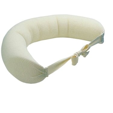
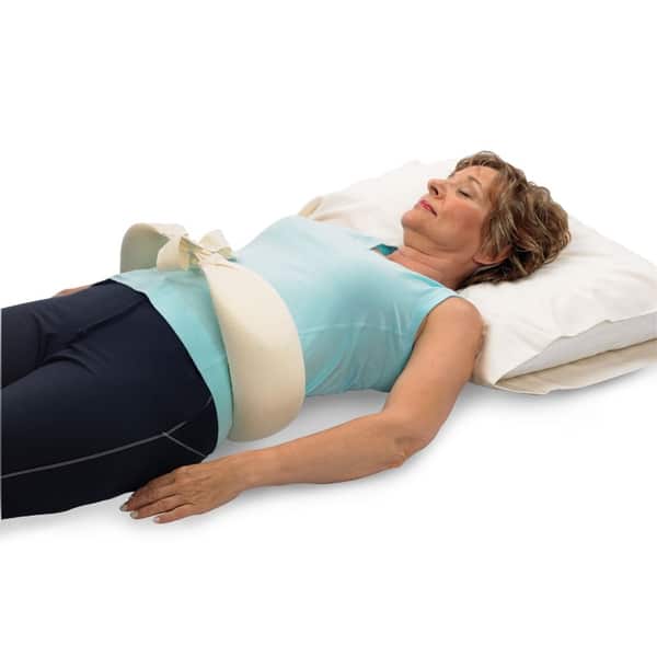
Original McKenzie Lumbar Night roll
In Stock
Price range: $55.95 through $69.95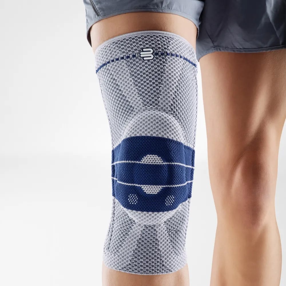
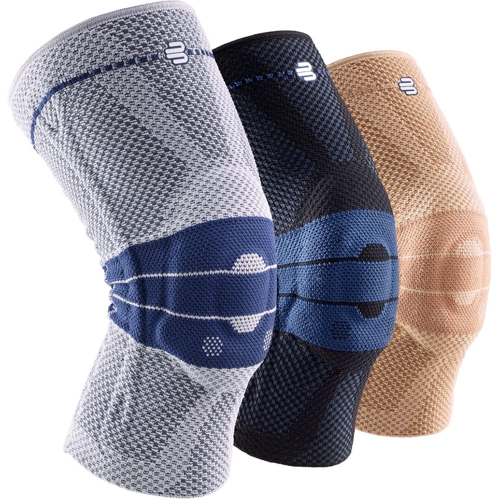
Bauerfeind GenuTrain Knee Brace (New Edition)
In Stock(2 Units)
$200.00


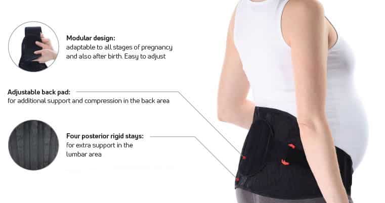
Donjoy Actistrap Back Support
In Stock
$119.95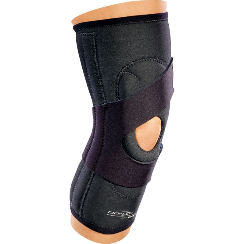

DonJoy Lateral J Patella Knee Brace
In Stock
$134.95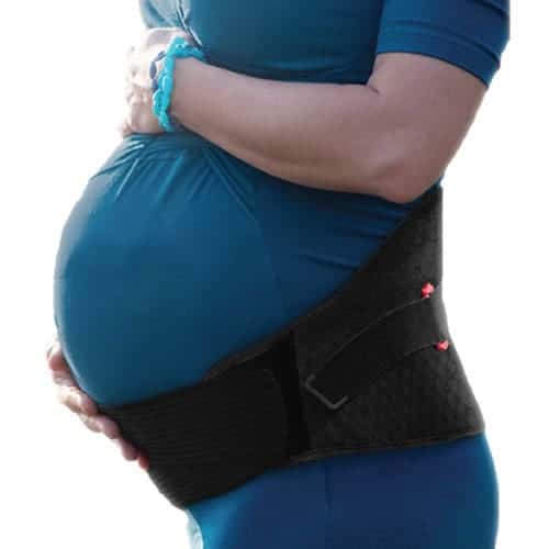
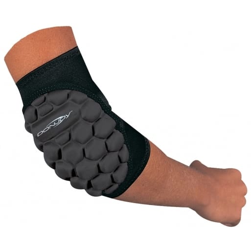
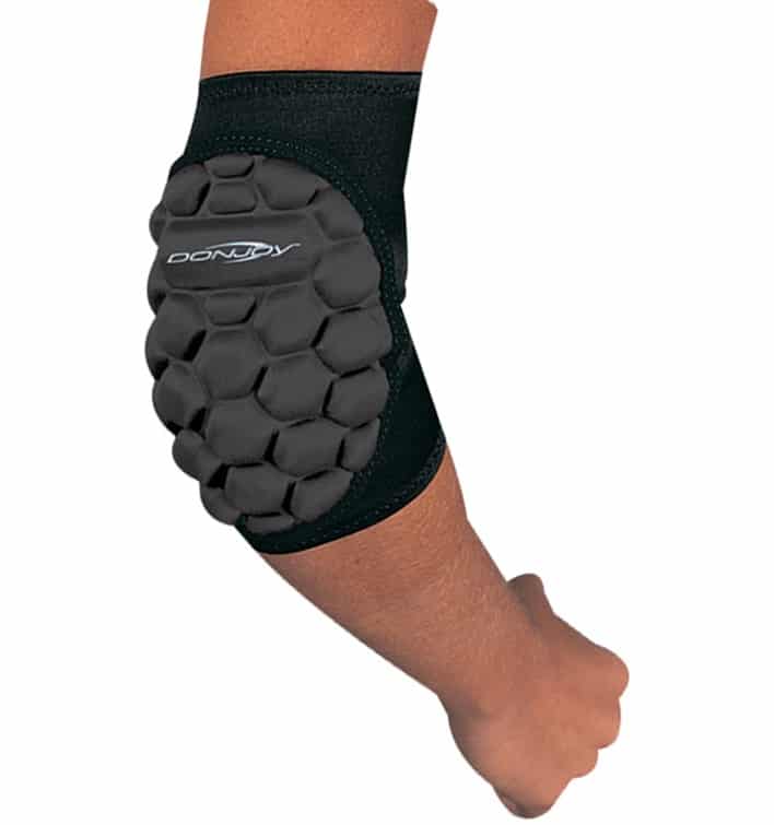
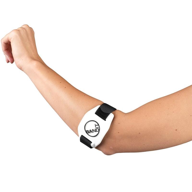
DonJoy Spider Elbow Pad
In Stock
$66.95
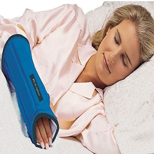
Procare Imak Pil-O-Splint – Wrist Night Splint
In Stock
$66.95
 SUPPORT SMALL CANADIAN BUSINESS


Reviews
There are no reviews yet