


TomLand Medical Life-Size 13-Part Anatomical Leg Muscle Model
Original price was: $749.95.$699.95Current price is: $699.95.
In stock (can be backordered)

Clinical Pricing Available!
The Physio Store is offering clinical pricing for all businesses / institutions in healthcare, healthcare professionals/students and fitness industry professionals.
We highly recommend you to sign up for our clinical pricing program, which will provide you with discounts on most of our products. We will also send out exclusive monthly specials, new ideas and new products on the market. Our company values the hard work and dedication of healthcare professionals and express gratitude by offering you special pricing and monthly deals.
Close

Manufacturer Warranty
2-Year Warranty
TomLand Medical anatomy models are covered by a 2-year warranty against manufacturing defects. This warranty applies to defects in materials or workmanship under normal educational, clinical or demonstration use.
Damage from misuse, dropping, improper handling, improper cleaning, modification, accidental damage or normal wear is not covered.
Return Policy
30-Day Return Window. Returns are accepted within 30 days of delivery for items that remain new and unopened.
Unopened condition required. Security seals must remain intact and packaging must not be opened. Items must be returned in original, unused condition for full product credit.
All law labels, identification labels, manuals, and original packaging must remain attached and undamaged. Please pack returns carefully in a secondary shipping box to prevent damage in transit.
If an item is returned opened, used, or not in resellable condition, the return will not be accepted. You will be contacted regarding return shipping fees to have the item shipped back to you.
If we do not receive instructions regarding return shipment within 30 days, you authorize us to dispose of the item on your behalf.
International orders are final sale. Orders shipped outside of Canada are not eligible for return or exchange.
For complete eligibility requirements and return instructions, please review our Return & Exchange Policy.

TomLand Medical Life-Size 13-Part Anatomical Leg Muscle Model
The TomLand Medical Life-Size 13-Part Anatomical Leg Muscle Model is a deluxe teaching model for studying the muscles of the human leg. It shows the superficial and deeper muscle layers of the lower extremity, along with vascular structures, nerves and ligaments.
This model is useful for clinics, classrooms and healthcare training settings. It gives clinicians, educators and students a clear way to explain leg muscle anatomy, muscle relationships, lower limb structure and movement.
Detailed Lower Extremity Muscle Anatomy
- Life-size anatomical leg model showing the muscles of the human leg
- Shows superficial and deeper muscle structures
- Includes vascular structures, nerves and ligaments
- Includes removable muscle parts for hands-on learning
- Useful for clinics, classrooms and healthcare training programs
Why Use a Life-Size Anatomical Leg Muscle Model?
Many students and patients find muscle anatomy hard to understand from diagrams alone. However, a three-dimensional life-size model makes it easier to see how the muscles of the leg relate to the bones, joints, nerves and vessels.
For example, an educator can remove selected muscles to show deeper structures. As a result, students can better understand how the superficial and deep layers of the lower extremity fit together.
Useful for Clinical Education and Hands-On Teaching
This anatomical leg model is well suited for teaching lower limb anatomy in professional and educational settings. It can support lessons on muscle location, muscle function, movement, injury patterns and rehabilitation goals.
In addition, healthcare professionals can use the model during patient education. It may help explain where a muscle, tendon or nerve-related concern is located in the leg.
Common Uses
- Teaching lower extremity muscle anatomy
- Explaining superficial and deep leg muscles
- Showing the relationship between muscles, nerves, vessels and ligaments
- Supporting physiotherapy, massage therapy and healthcare education
- Clinical demonstration in treatment rooms, classrooms and training labs
Removable Parts
This 13-part model includes removable muscle and lower limb structures for more detailed teaching. The removable parts include:
Removable Anatomy Structures
- Sartorius
- Long head of biceps femoris
- Gluteus maximus
- Soleus
- Gastrocnemius
- Gracilis
- Semitendinosus and semimembranosus
- Rectus femoris
- Extensor digitorum longus
- Sole of the foot
- Tensor fasciae latae
Anatomy Shown
The TomLand Medical Life-Size 13-Part Anatomical Leg Muscle Model shows detailed lower limb anatomy. It represents the muscles of the leg, including both surface-level and deeper structures.
It also shows important supporting structures such as nerves, blood vessels and ligaments. Therefore, it is useful for teaching how the lower limb is organized and how different structures relate to movement, support and function.
| Product Type | Life-size anatomical leg muscle model |
|---|---|
| Model Format | 13-part lower extremity muscle model |
| Scale | Life-size |
| Region Shown | Human leg / lower extremity |
| Anatomy Included | Superficial muscles, deeper muscles, nerves, vascular structures and ligaments |
| Removable Parts | Includes removable muscle and foot structures for hands-on anatomy education |
| Pathology | No pathology shown |
| Use | Clinical education, anatomy teaching, classroom training and patient demonstration |
| Suitable For | Clinics, classrooms, healthcare training programs and professional education settings |
| Brand | TomLand Medical |
Helpful for Explaining Leg Muscles and Movement
This model can help explain how major leg muscles support movement at the hip, knee, ankle and foot. It can also help show how muscle groups work together during standing, walking, running and rehabilitation exercises.
Because the model includes removable parts, it can support step-by-step teaching. For example, an educator can start with the surface muscles and then remove selected parts to show deeper anatomy.
Helpful Discussion Topics
- Superficial and deep muscles of the human leg
- Muscle relationships around the hip, knee, ankle and foot
- Basic lower limb movement and support
- How muscles, nerves, vessels and ligaments relate to one another
- Lower extremity anatomy for students, clinicians and patient education
Who Uses This Leg Muscle Model?
This model is useful for healthcare professionals, educators and students who need a clear way to teach lower extremity anatomy. It works well in physiotherapy clinics, massage therapy programs, chiropractic offices, medical offices, rehabilitation centres, classrooms and healthcare training programs.
Ideal For
- Physiotherapists
- Massage therapists
- Chiropractors
- Physicians and medical offices
- Healthcare educators and students
- Schools, universities and clinical training programs
TomLand Medical Clinical Education Products
TomLand Medical products are designed for practical use in professional healthcare settings. This life-size 13-part anatomical leg muscle model gives clinics and educators a detailed visual tool for teaching lower extremity muscle anatomy.
Available in Canada
TomLand Medical products are available to clinics, healthcare professionals, schools, universities, rehabilitation programs and training organizations across Canada.
Frequently Asked Questions
What does the TomLand Medical Life-Size 13-Part Anatomical Leg Muscle Model show?
This model shows the muscles of the human leg, including superficial and deeper muscle structures. It also represents vascular structures, nerves and ligaments.
Is this anatomical leg model life-size?
Yes. This is a life-size anatomical leg muscle model, which makes it useful for detailed teaching, clinical demonstration and hands-on anatomy education.
Is this anatomical leg model a 13-part model?
Yes. This is a 13-part anatomical leg muscle model with removable parts for hands-on teaching and demonstration.
What removable parts are included?
Removable parts include the sartorius, long head of biceps femoris, gluteus maximus, soleus, gastrocnemius, gracilis, semitendinosus and semimembranosus, rectus femoris, extensor digitorum longus, sole of the foot and tensor fasciae latae.
Does this leg model show nerves and blood vessels?
Yes. The model represents nerves and vascular structures, making it useful for teaching how muscles, vessels and nerves relate to one another in the lower extremity.
Does this model show pathology?
No. This model shows normal lower extremity anatomy. It is designed for anatomy education and demonstration, not for showing disease or injury changes.
Who would use this anatomical leg muscle model?
This model is useful for physiotherapists, massage therapists, chiropractors, physicians, healthcare educators, students, rehabilitation clinics and training programs.
Is this model useful for massage therapy education?
Yes. Massage therapy programs can use this model to teach leg muscle anatomy, muscle location, muscle layers and the relationship between superficial and deeper structures.
Is this model useful for physiotherapy education?
Yes. Physiotherapy clinics and training programs can use this model to explain lower limb muscles, movement, rehabilitation goals and basic lower extremity anatomy.
Can this model be used for patient education?
Yes. Clinicians can use this model to help patients understand where muscles, tendons, nerves or related lower limb structures are located.
Can this model be used in schools or healthcare training programs?
Yes. This model is suitable for colleges, universities, healthcare classrooms, clinical skills labs and professional training programs.
What is the benefit of removable muscle parts?
Removable muscle parts allow educators and students to study surface anatomy first, then remove selected structures to see deeper layers and relationships.
Is this anatomical leg model a diagnostic tool?
No. This model is for education and demonstration only. It can help explain anatomy, but it does not replace assessment, diagnosis or treatment from a qualified healthcare professional.
| Country of Origin | China |
|---|---|
| HS Code | 9023.00.00.00 |
| Availability |
Now available in-store at 22635 Komoka Road Komoka N0L1R0 Powered With Oliver Pos |
|---|
What does the TomLand Medical Life-Size 13-Part Anatomical Leg Muscle Model show?
This model shows the muscles of the human leg, including superficial and deeper muscle structures. It also represents vascular structures, nerves and ligaments.
Is this anatomical leg model life-size?
Yes. This is a life-size anatomical leg muscle model, which makes it useful for detailed teaching, clinical demonstration and hands-on anatomy education.
Is this anatomical leg model a 13-part model?
Yes. This is a 13-part anatomical leg muscle model with removable parts for hands-on teaching and demonstration.
What removable parts are included?
Removable parts include the sartorius, long head of biceps femoris, gluteus maximus, soleus, gastrocnemius, gracilis, semitendinosus and semimembranosus, rectus femoris, extensor digitorum longus, sole of the foot and tensor fasciae latae.
Does this leg model show nerves and blood vessels?
Yes. The model represents nerves and vascular structures, making it useful for teaching how muscles, vessels and nerves relate to one another in the lower extremity.
Does this model show pathology?
No. This model shows normal lower extremity anatomy. It is designed for anatomy education and demonstration, not for showing disease or injury changes.
Who would use this anatomical leg muscle model?
This model is useful for physiotherapists, massage therapists, chiropractors, physicians, healthcare educators, students, rehabilitation clinics and training programs.
Is this model useful for massage therapy education?
Yes. Massage therapy programs can use this model to teach leg muscle anatomy, muscle location, muscle layers and the relationship between superficial and deeper structures.
Is this model useful for physiotherapy education?
Yes. Physiotherapy clinics and training programs can use this model to explain lower limb muscles, movement, rehabilitation goals and basic lower extremity anatomy.
Can this model be used for patient education?
Yes. Clinicians can use this model to help patients understand where muscles, tendons, nerves or related lower limb structures are located.
Can this model be used in schools or healthcare training programs?
Yes. This model is suitable for colleges, universities, healthcare classrooms, clinical skills labs and professional training programs.
What is the benefit of removable muscle parts?
Removable muscle parts allow educators and students to study surface anatomy first, then remove selected structures to see deeper layers and relationships.
Is this anatomical leg model a diagnostic tool?
No. This model is for education and demonstration only. It can help explain anatomy, but it does not replace assessment, diagnosis or treatment from a qualified healthcare professional.
You may also like…


TomLand Medical Advanced Colour Spine Model with Muscles & Disc Herniation Demo
In Stock(1 Units)
$299.95


Related products
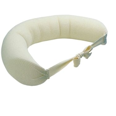
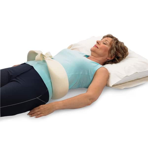
Original McKenzie Lumbar Night roll
In Stock
Price range: $55.95 through $69.95

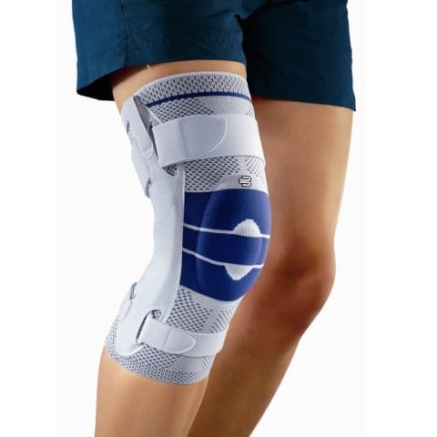
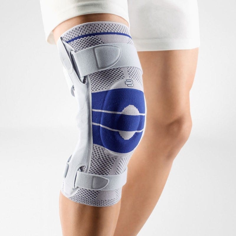
Bauerfeind GenuTrain S Knee Brace
In Stock
$400.00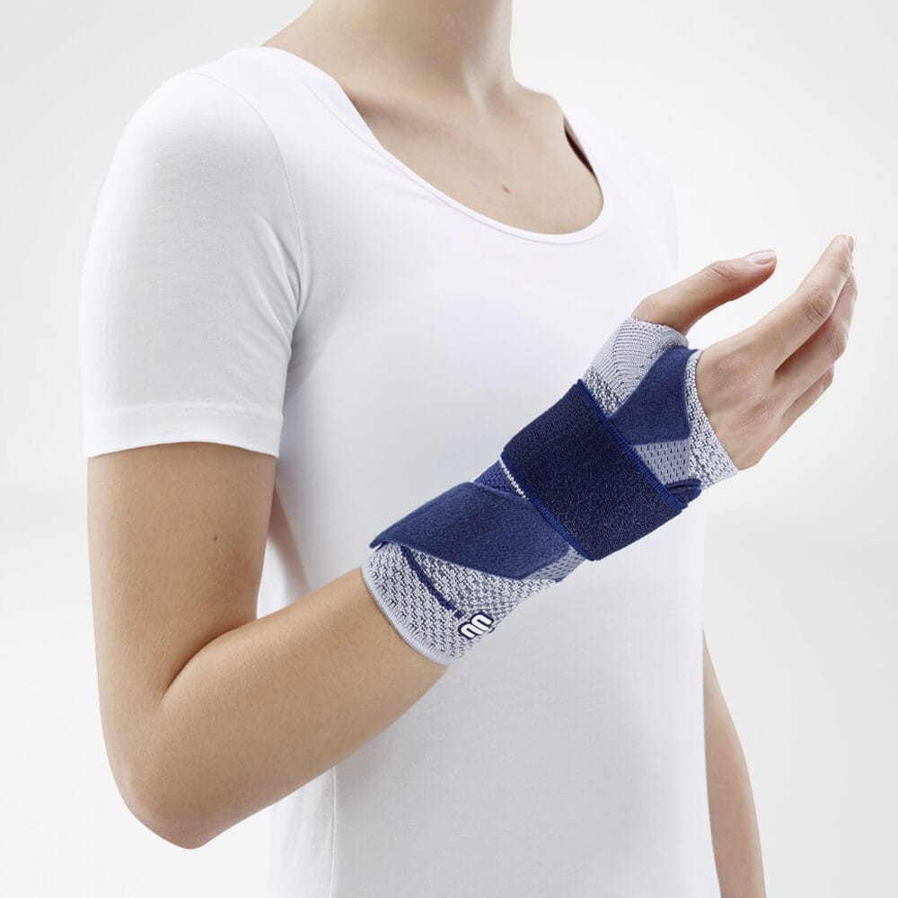
Bauerfeind ManuTrain Wrist Brace
In Stock
$195.00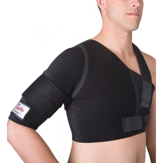
DonJoy Sully® Shoulder Stabilizer Brace
In Stock
$349.95

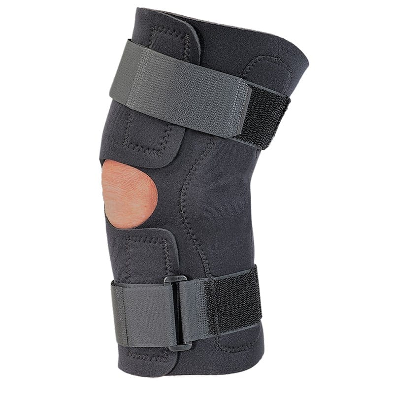
MKO Hinged Knee Wrap
In Stock
$89.99
 SUPPORT SMALL CANADIAN BUSINESS